Welcome to the 3D Brachial Plexus MRI/MRA/MRV Image Gallery
[images courtesy of Dave Nelson and Ray Guerrero of the UCLA Radiology Media Center, annotated by James D. Collins, MD (UCLA Radiology)]
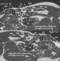
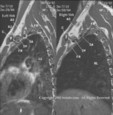
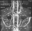
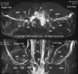
These are images of a person diagnosed with bilateral Thoracic Outlet Syndrome (TOS). Click on annotated image to see a larger view. Please check back regularly as descriptions of the images are being added (and additional images will be added in the future).
The radiologist interprets results from the monitored 3D MRI/MRA/MRV by viewing sequential images at the imaging console. The 2D Time-of-Flight (TOF) MRA/MRV (angiography/venography) sequence images the vascular supply in high resolution. Software allows the 2D TOF MRA/MRV sequence to be viewed as a rotating 3D image on the console. Sequences of closely-spaced 2D high-resolution MRI at 4.0 mm are obtained in 5 planes: coronal, transverse, transverse oblique, sagittal, and coronal abduction external rotation of the upper extremities (AER). Software interpolation allows 3D image reconstruction for the radiologist to review and annotate the images. Chest X-rays are obtained for landmark anatomy and to evaluate bony abnormalities (e.g., cervical rib, elongated transverse process of C7, clavicular fractures, scoliosis, rounding of the shoulders, and scarring within soft tissues). A detailed description of Dr. Collins's 3D MRI/MRA/MRV technique is reprinted with his permission.
The 3D MRI/MRA/MRV technique was published in the
April 2003 edition of the Journal of the National Medical Association (Vol 95:298-306).
Compromising abnormalities of the brachial plexus displays the blood supply to a nerve model, 3D mass effect from silicone rupture, thyroid goiter, fractured clavicle, body builder (enlarged muscles), and anomalous muscle that triggered complaints of thoracic outlet syndrome. Published online January 24th, 2005 Clinical Anatomy volume 8 issue 1, pages 1-16 Wiley-Liss.
The most important message from this display is to know anatomy. Image an artery and you image a nerve. Nerves DO have a blood supply. "You only see what you know."
A 52-minute QuickTime video featuring Dr. Collins interpreting a 3D brachial plexus MRI/MRA/MRV of a patient with bilateral Thoracic Outlet Syndrome (TOS) can be downloaded here.
Return to tosinfo HOME PAGE
Comments or suggestions? Contact the webmaster at stevedo@gmail.com.
[Last update: Wednesday March 25th, 2009]