Return to IMAGE GALLERY
These are selected images from the sagittal sequence. In
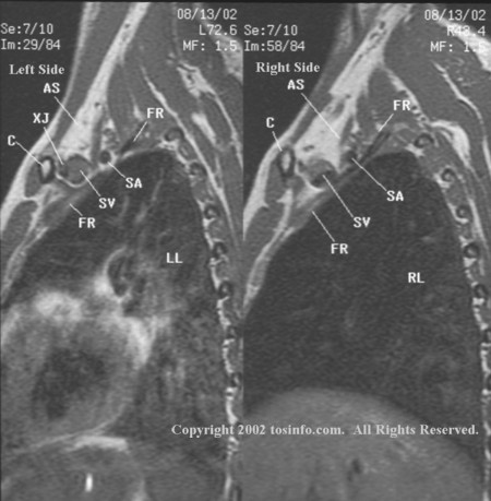
the sagittal sequence, images are perpendicular to the line of sight
when viewing the supine patient (with arms at the sides) from the shoulder
toward the opposite shoulder.
The sagittal sequence cross-references the coronal and
transverse
sequences to display costoclavicular compression of the external jugular vein (XJ) against the bicuspid valve within the subclavian vein (SV) on
the first rib (FR), costoclavicular compression of the axillary
vein against the artery, interscalene triangle compression, and scarring
(fibrosis) when present. The left and right
sagittal images were selected to display the close proximity of the
clavicle (C) to the external jugular (XJ) and subclavian veins (SV),
particularly on the left. When the arms are elevated above the head, the
posterior inferior rotation of the clavicles with the subclavius muscles
efface and/or compress the venous drainage of the upper extremities.
In these neutral images, the gray proton density (color) within the left subclavian vein
(SV) reflects decreased venous flow as compared to the right subclavian vein.
A few labels have been placed on the image to assist with identification
of landmark anatomy. The clavicles (C), external jugular vein (XJ),
right lung (RL), left lung (LL), first ribs (FR), subclavian veins (SV),
subclavian arteries (SA), and anterior scalene muscles (AS) are labeled
for reference.